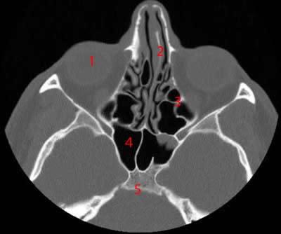
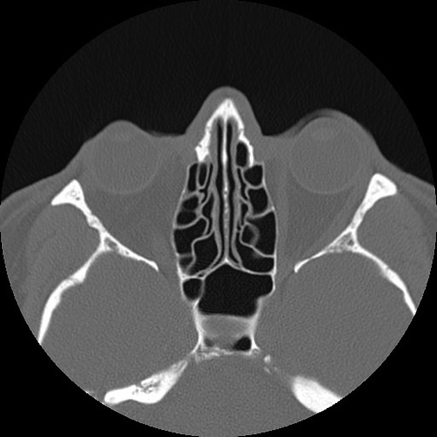
[最も好ましい] sinuses labeled on ct 281322-Sinuses labeled on ct
Mar 01, 09 · SUMMARY Our aim was to review the imaging findings of relatively common lesions involving the cavernous sinus (CS), such as neoplastic, inflammatory, and vascular ones The most common are neurogenic tumors and cavernoma Tumors of the nasopharynx, skull base, and sphenoid sinus may extend to the CS as can perineural and hematogenous metastasesMay 18, 21 · CT anatomy of the nasal cavity and paranasal sinuses is described in detail together with the anatomic variants encountered in each region Imaging techniques and protocol Preoperative CT imaging of the paranasal sinuses is performed after completion of the medical treatment because up to 80% of patients suffering from acute upper respiratory tract infectionOct 13, 14 · CT anatomy of the paranasal sinuses 1 1 Paranasal Sinuses Radiological Anatomy Dr Hazem Abu Zeid Youset December 06 2 2 What are the sinuses?

Paranasal Sinuses Annotated Ct Radiology Case Radiopaedia Org
Sinuses labeled on ct
Sinuses labeled on ct-Patients with facial trauma, positive paranasal sinus pathology, head and neck tumours and previous surgery were excludedIn the past decade in particular, CT of the paranasal sinuses has become a roadmap for FESS The radiologist's goal is to report on five key points the extent of sinus opacification, opacification of sinus drainage pathways, anatomical variants, critical variants, and condition of surrounding soft tissues of the neck, brain and orbits


Sinusitis Acute
Aug 08, 18 · In addition to reviewing the scan to determine the presence of disease, CT scans of the sinuses can also be reviewed to evaluate potential areasComputed tomography (CT) of the sinuses uses special xray equipment to evaluate the paranasal sinus cavities – hollow, airfilled spaces within the bones of the face surrounding the nasal cavity CT scanning is painless, noninvasive and accurate It's also the most reliable imaging technique for determining if the sinuses are obstructed andFeb 19, 21 · MC Huguelet A sinus CT scan is performed to produce images of the sinuses A sinus CT scan is an imaging test that uses advanced xray technology to produce a detailed image of the sinuses, hollow passages of indeterminate function that are found within the skull Typically, the purpose of a sinus CT scan is to aid a doctor in diagnosing a medical condition involving the sinuses
Nov 30, 16 · Computed tomography (CT) is considered to be a radiological method that can evaluate the anatomy and anatomical variants of the paranasal sinuses in the right way and is extremely useful in theNumerous sinonasal anatomic variants exist and are frequently seen on sinus CT scans The most common ones are Agger nasi cells, infraorbital ethmoidal (Haller) cells, sphenoethmoidal (Onodi) cells, nasal septal deviation, and concha bullosa 1–10The Agger nasi cells are the most anterior ethmoidal air cellsThe ethmoid sinuses also called ethmoid labyrinth are located between the eyes and the nose Sinus in anatomy a hollow cavity recess or pocket In bones behind your nose are your sphenoid sinuses There are four pairs of paranasal sinuses the frontal sinuses are located above the eyes in the forehead bone The sphenoid sinuses are
• Standard coronal CT is INADEQUATE to understand 3D sinus anatomy, particularly in the frontal recess • Axial thin section (Mar 01, 17 · The patients were referred for computed tomography (CT) scan due to a clinical symptoms referable to sinonasal region A total of 365 consecutive CT studies of the paranasal sinuses were identified;Welcome to Interactive CT Sinus Anatomy Imaging the paranasal sinuses is routine in clinical practice to evaluate for various sinus pathology, nonspecific facial pain, and preoperative planning for functional endoscopic sinus surgery (FESS), including postoperative followup Our goal is to review the complex sinonasal anatomy, anatomic variants, mucociliary drainage pathways and inflammatory sinus
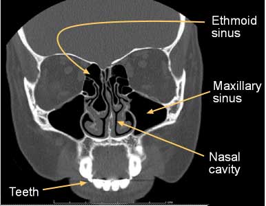


Computed Tomography Ct Sinuses
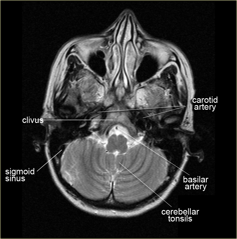


The Radiology Assistant Anatomy
The present work aimed to describe the normal computed tomography (CT) and crosssectional anatomy of the nasal and paranasal sinuses in sheep and to correlate these features with the relevant clinical practices Twenty apparent healthy heads of Egyptian native breed of sheep (Baladi sheep) of bothFeb 15, 21 · Unlike CT scans, MRIs do not show bone, so details about the bony walls and anatomy of the sinuses shown on CT are not visible with MRI Even the OMC is not easily seen MRIs are excellent for delineating soft tissue, so they can be useful in the rare case of a suspected sinusI want to work through the anatomy of the head as seen on CT imaging sections, but there's a lot to look at Let's start by seeing if we can recognise the fr
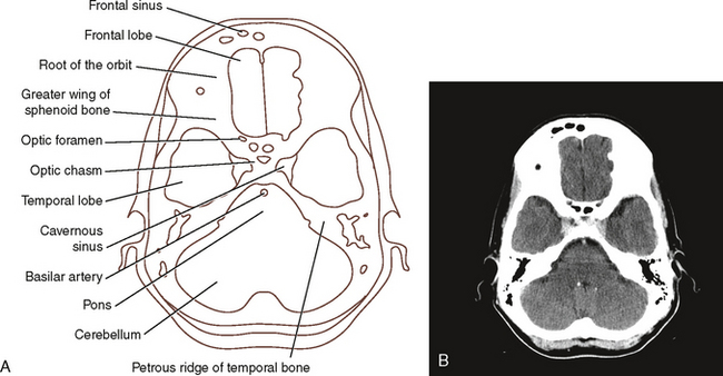


Sectional Anatomy For Radiographers Radiology Key


Pin On Anatomia
Feb 25, 09 · The CT clearly shows the opacified sinus, which is slightly hyperdense The signal characteristics on MRI and the attentuation on CT are a result of the high protein content of fungus This is a good example of the pitfall of the 'pseudopneumatized sinus'7 public playlist include this case A/P by Alexa Manickas Labeled Anatomy by Abdullah Abohimed IMPORTANTS by Dr Ahmed Faiz AlMusawi Annotated CTs by Reid Cline anatomi by mehmet işçi Annotated teaching by Doctor Murat Dzhanibekov LearningSectionalAnatomy by Dr Payam RiahiIn many ways CT scanning works very much like other xray examinations Xrays are a form of radiation—like light or radio waves—that can be directed at the body Different body parts absorb the xrays in varying degrees


Ct Scan Of The Sinuses Coronal View At The Level Of The Sphenoid Download Scientific Diagram
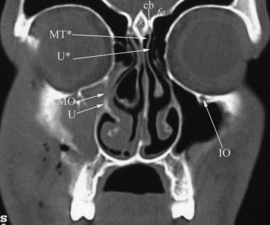


Ct Scan Of The Paranasal Sinuses History Basic Concepts Anatomy
CT scans are one of the safest ways to study the sinuses, and they offer the most reliable imaging for diagnosing obstructions and other problems What to expect from a CT scan of the sinuses Having a CT scan of your sinuses is a simple procedure that takes place at our officeCt sinuses anatomy Other functions are air humidification and aiding in voice resonance Imaging the paranasal sinuses is routine in clinical practice to evaluate for various sinus pathology non specific facial pain and pre operative planning for functional endoscopic sinus surgery fess including post operative follow up The ct test isJul 01, 19 · Sphenoid sinuses CT brain (bone windows) The sphenoid sinus can be single or septated into multiple separate sinuses Sphenoid sinus clinical significance In the context of trauma, a fluid level in the sphenoid sinus may be a sign of a basal skull fracture


Head And Neck Undergraduate Diagnostic Imaging Fundamentals


Www Mdpi Com 75 4418 11 2 250 Pdf
Mar 14, 10 · There are four sets of sinuses maxillary, ethmoid, frontal and sphenoid sinuses We will examine most of them in the following series of drawings and CT scans The initial concepts are a little difficult to understand, but will become clearer when we get to the CT scansThe sinuses are chambers in the bones of the face and skull that are normally lined with a thin mucosa They communicate with the nasal cavity via narrow openingsComputed Tomography (CT) Sinuses2 May 23, 07 How does the procedure work?


Ct Angio Atlas Neuroangio Org


Skull Base Related Lesions At Routine Head Ct From The Emergency Department Pearls Pitfalls And Lessons Learned Radiographics
For a ct scan of the sinuses the patient is most commonly positioned lying flat on the back The paranasal sinuses usually consist of four paired air filled spaces Welcome to interactive ct sinus anatomy Ct is currently the modality of choice in the evaluation of the paranasal sinuses and adjacent structuresA CT scan may help detect sinusitis, evaluate sinuses filled with thickened sinus membranes, give additional information on tumors of the nasal cavity, diagnose inflammatory disorders, and plan for surgery by defining anatomy(12) The preferred initial procedure is a coronal CT image(13)Nov 15, 02 · CT scans can provide much more detailed information about the anatomy and abnormalities of the paranasal sinuses than plain films12 A CT scan provides greater definition of the sinuses and is


Sinusitis Acute


Sectional Anatomy Study Tool Radiography
Sinus anatomy, the commonly used imaging technology in 1985, standard plain films and polytomography, was quickly replaced with CT Coronal CT scans afforded improved resolution of the bony framework and the superimposed mucosa in addition to regional inflammatory pathology The application of multiplanar reconstruction andNormal sinus anatomy is complex, and can be difficult to appreciate on static images alone Therefore, this exhibit contains an interactive atlas of the normal sinuses in the axial, sagittal and coronal planes As a demonstration, please move your cursor over the 3D skull below to scroll through a coronal stack of CT images Besides this interactive atlas, this exhibit also includesFeb 09, 21 · Anatomy of the head on a cranial CT Scan brain, bones of cranium, sinuses of the face Coronal Brain CT Vasculary territories Dural venous sinuses, Veins, Arteries Bones of cranium Axial CT Paranasal sinuses CT Cranial base , CT Foramina, Nasal cavity, Paranasal sinuses Bones of cranium Anatomy , CT Invalid input


File Ct Scan Of A Pacchionian Body Transverse Plane Labeled Jpg Wikimedia Commons


Brain Ct Anatomy شفا عظیم عصبي جراحي اختصاصي روغتون Facebook
Aug , 19 · The turbinates and meati are best seen on coronal CT sections ( Fig 231 ) The middle turbinate and meatus form the most important anatomic area in the lateral wall of the nose The attachments of the middle turbinate have anatomic and surgical relevance and provide fairly good stability to the turbinateJul 26, 16 · 43 Frontal Sinus 44 Inner Ear 45 Sigmoid Sinus 46 Internal Carotid Artery 47 Sphenoid Bone 48 Sphenoid Sinus 49 Medulla Oblongata 50 External Auditory Meatus 51A good knowledge of the complex CT anatomy of the paranasal sinuses is crucial This knowledge will provide an accurate assessment of the normal variants and pathological changes


Maxillary Sinus Radiology Reference Article Radiopaedia Org
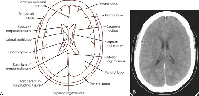


Sectional Anatomy For Radiographers Radiology Key
Jan 09, 11 · 76 Fundamentals of Sectional Anatomy CT IMAGES Exam 1 Coronal Images Frontal bone Frontal sinus Perpendicular plate of ethmoid bone Nasal bone Frontal process of maxillary bone Figure 213 Although Figure 213 is not the first image of this exam, it is anterior enough to demonstrate the unusually large but typical asymmetric frontal sinusesTheDescription This workshop on paranasal sinuses will focus on detailed CT anatomy and important paranasal sinus landmarks You will learn how to prospectively identify important normal variants that predispose patients for surgical complications and how to easily navigate through the complex structures of a PNS CTSIMPLIFIED PARANASAL ANATOMY #anatomy #pns #ctpns Also known as antrum of Highmore Largest paranasal sinusPyramidal in shape Base toward the lateral wall of


Dural Venous Sinuses 3d Anatomy Tutorial Youtube


Great Vessel And Coronary Artery Anatomy In Transposition And Other Coronary Anomalies A Universal Descriptive And Alphanumerical Sequential Classification Sciencedirect
Labeled imaging anatomy cases Dr Calum Worsley and Assoc Prof Craig Hacking et al This article lists a series of labeled imaging anatomy cases by system and modality On this page Article Brain Head and neck Chest Abdomen and pelvisMay 16, 19 labelled CT sinus coronal Google SearchSearch from Sinuses Anatomy Pictures stock photos, pictures and royaltyfree images from iStock Find highquality stock photos that you won't find anywhere else


Imaging Of Maxillary Sinusitis Waters View Ct Scan Mri Otolaryngology Houston
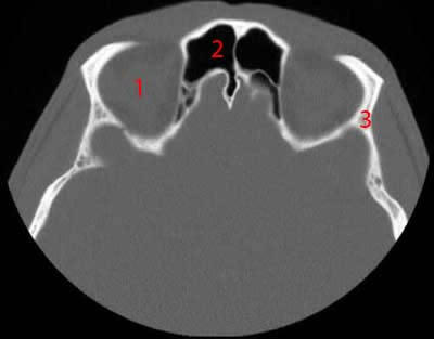


Paranasal Sinuses Ct Anatomy W Radiology
Sinus The neurovascular anatomy of the cavernous sinus is reviewed and correlated with CT findings Discrepancies between anatomic and CT appearance are discussed The cavernous sinus is a unique component of the cranial vascular system, having direct or indirect connections with the cerebrum, cerebellum, brainstem,Nov 11, 19 · #2 Anatomy of the Paranasal Sinuses This excellent 3D model uploaded b y valchanov shows Maxillary sinus one sinus located within the bone of each cheek Ethmoid sinus located under the bone of the inside corner of each eye, although this is often shown as a single sinus in diagrams, this is really a honeycomblike structure of 612 small sinuses that is better appreciated on CTSep 19, 16 · An additional important sinus drainage pathway that may be targeted with FESS is the frontal recess, which is best evaluated on sagittal reformatted CT images and is located along the posterior margin of the agger nasi cell (most anterior ethmoid air cell) or other variants of frontal recess cells, when present (Fig 2) (10)


Ct Scan Of Paranasal Sinus Axial Coronal Youtube


Ent Radiological Anatomy
Sep 09, 09 · Computed tomography ( CT scan ) A CT scanner uses Xrays and a computer to create detailed images of the sinuses CT scanning can help diagnose chronic sinusitis Magnetic resonance imaging ( MRIJan 13, 15 · Understand your individual sinus anatomy In some cases of chronic sinus infections, surgery is an option to remove tissue or a polyp if that's blocking a nasal or sinusAJR194, June 10 W529 Multiplanar Sinus CT A C E Fig 1—34yearold woman with normal sinus anatomy A, Coronal CT image shows ostiomeatal complex, which consists of four structures hiatus semilunaris (oval), uncinate process (arrowhead), infundibulum (dotted line), ethmoidal bulla (EB), and maxillary ostium (asterisk)Max = maxillary sinus, IT = inferior turbinate, MT = middle


Ct Sinuses Anatomy Quiz Radiology Case Radiopaedia Org


Paranasal Sinuses Annotated Ct Radiology Case Radiopaedia Org
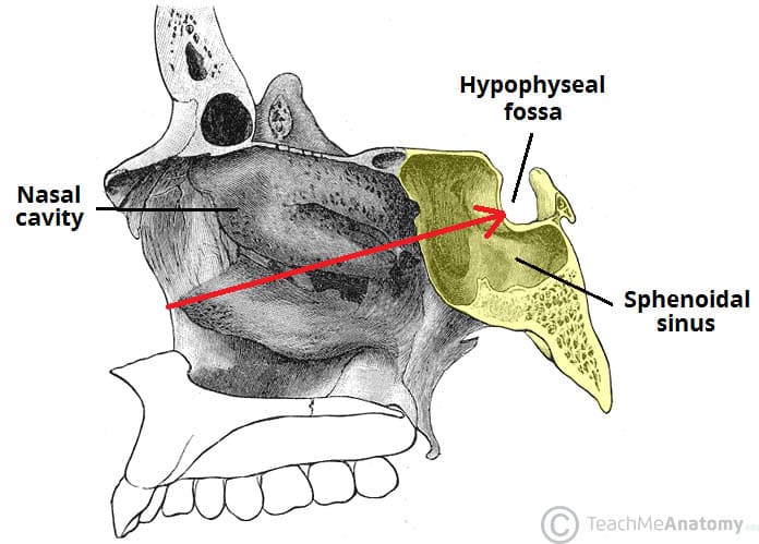


The Paranasal Sinuses Structure Function Teachmeanatomy


Paranasal Sinuses Maxillary Ethmoid Sphenoid Frontal Notes Youtube


What Is The Significance Of Absent Frontal Sinuses Does This Hinder The Way Sinuses Are Supposed To Work What Effect Does Missing Frontal Sinus Have On A Person Quora
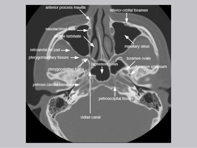


Cross Sectional Anatomy Of Paranasal Sinus


Approach To Ct Head Learningneurology Com


1


Startradiology


Transverse Computed Tomography Image Of The Head Warmblood Stallion Download Scientific Diagram


A Ct Scan In Coronal Plane Shows The Normal Paranasal Sinus Anatomy Download Scientific Diagram


Mri Neck Anatomy Free Mri Axial Neck Cross Sectional Anatomy
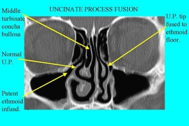


Nasal Cavity Anatomy Physiology And Anomalies On Ct Scan Overview Anatomy And Physiology Of The Nasal Cavity Nasal Cavity Anomalies And Sinusitis


Ct Scan Pns Coronal Cuts Ct Scan Machine


1


Onlinelibrary Wiley Com Doi Pdf 10 1097


Ct Scan Anatomy Of Paranasal Sinuses Professor Dr Muhammad Ajmal Ppt Video Online Download


Radiology Of The Nasal Cavity And Paranasal Sinuses Ento Key


Cavernous Sinus And Its Cranial Nerves


Approach To Ct Head Learningneurology Com


Head Phantom


Q Tbn And9gcthldxvr6nykftmuawhyfamnfge8pfsjllwgh3teyo Usqp Cau


Plos One Bold Granger Causality Reflects Vascular Anatomy
:background_color(FFFFFF):format(jpeg)/images/library/12298/mri-t2-axial-caudate-nucleus-level_english.jpg)


Radiological Anatomy X Ray Ct Mri Kenhub
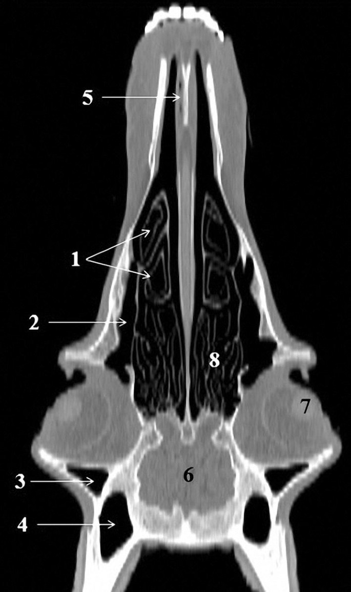


Anatomy Of The Head In The Saanen Goat A Computed Tomographic And Cross Sectional Approach Springerlink
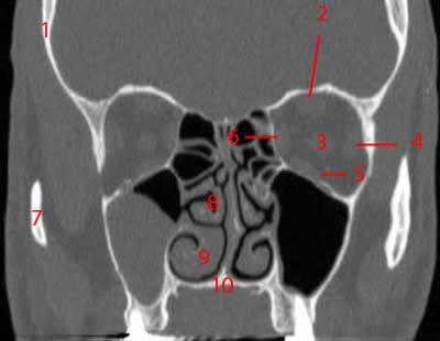


Paranasal Sinuses Ct Anatomy W Radiology
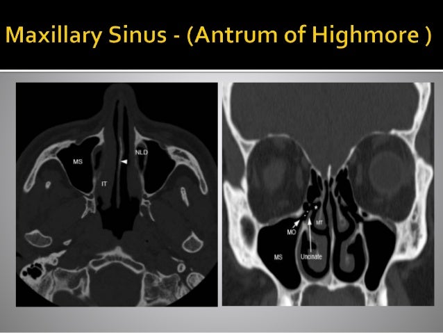


Ct Anatomy Of Para Nasal Sinuses


Radiology Of The Nasal Cavity And Paranasal Sinuses Ento Key


Mrv Major Venous Sinuses Are Labeled With Arrows Download Scientific Diagram


Brain And Face Ct Interactive Anatomy Atlas


Radr 2331 Parietoacanthial Projection Waters Method Sinuses Anatomy Diagram Quizlet
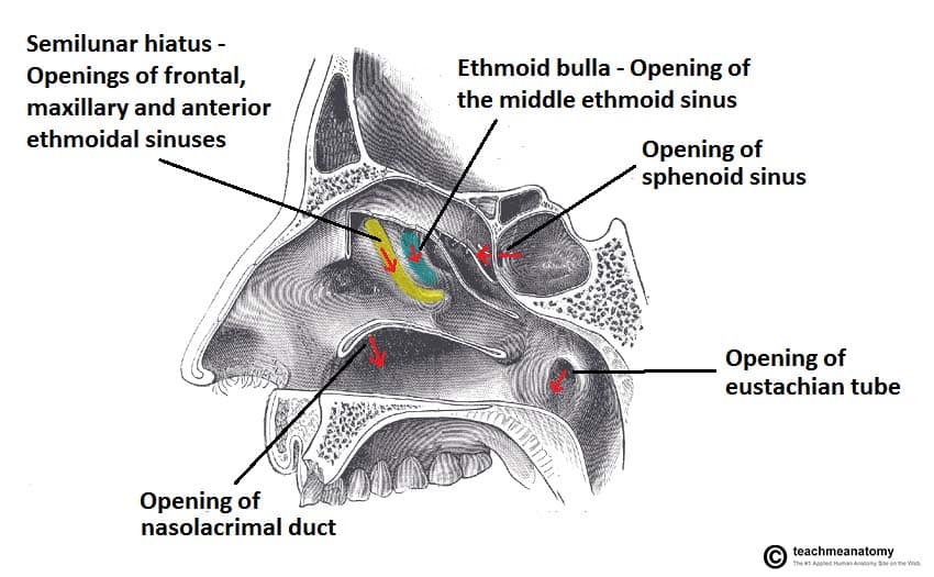


The Paranasal Sinuses Structure Function Teachmeanatomy


Onlinelibrary Wiley Com Doi Pdf 10 1097
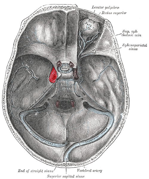


Cavernous Sinus Wikiwand


Ct Anatomy Brain Anatomy Drawing Diagram


Approach To Ct Head Learningneurology Com
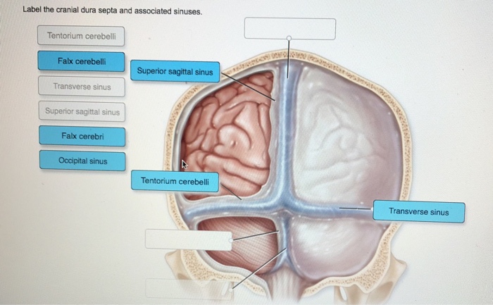


Label The Cranial Dura Septa And Associated Sinuses Chegg Com


Transverse Sinuses Wikipedia


Ct Scan Of Head And Neck


Sinusitis Mastoiditis Undergraduate Diagnostic Imaging Fundamentals
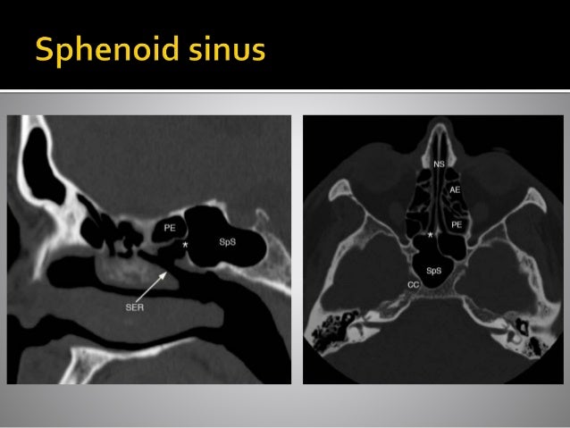


Ct Anatomy Of Para Nasal Sinuses


Computed Tomography Anatomy Of The Paranasal Sinuses And Anatomical Variants Of Clinical Relevants In Nigerian Adults Sciencedirect


Imaging Lesions Of The Cavernous Sinus American Journal Of Neuroradiology


Ent Radiological Anatomy


File Dural Sinuses Jpg Wikimedia Commons


Paranasal Sinuses Ct Anatomy W Radiology
:background_color(FFFFFF):format(jpeg)/images/library/12299/axial-ct-jugular-fossa-level_english.jpg)


Radiological Anatomy X Ray Ct Mri Kenhub


Home Page


Startradiology


Startradiology


Front View Illustration And Side By Side Ct Scans Of Normal And Chronic Sinusistis Labeled Frontal Sinus Ethmoid Sinuses Maxillary Sinus Nasal Septum Eye Socket Sinusitis Is Inflammation Of The Paranasal Sinuses Which May


Module 1 2 Flashcards Quizlet


Normal Sinus Ct Annotated Radiology Case Radiopaedia Org


Nose Anatomy And Histology Of The Human Nose Medical Library


9 Best Sinus Diagram Images Stock Photos Vectors Adobe Stock


Imaging Spectrum Of Cavernous Sinus Lesions With Histopathologic Correlation Radiographics


Onlinelibrary Wiley Com Doi Pdf 10 1111 J 1740 61 00 Tb X
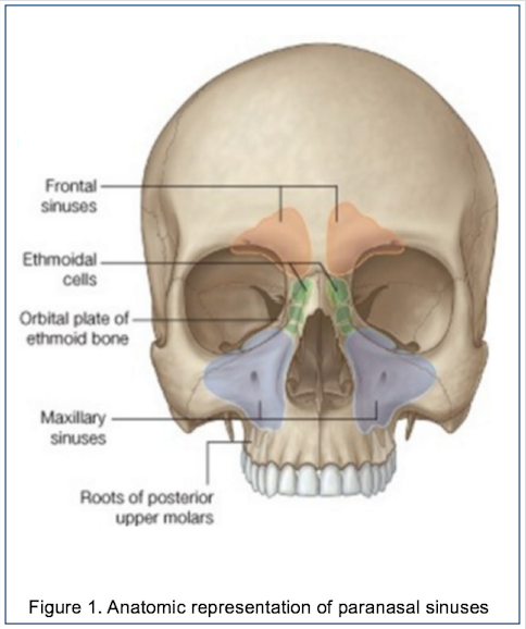


Epos C 2117


Imaging Lesions Of The Cavernous Sinus American Journal Of Neuroradiology


Radiology Basics Head Anatomy
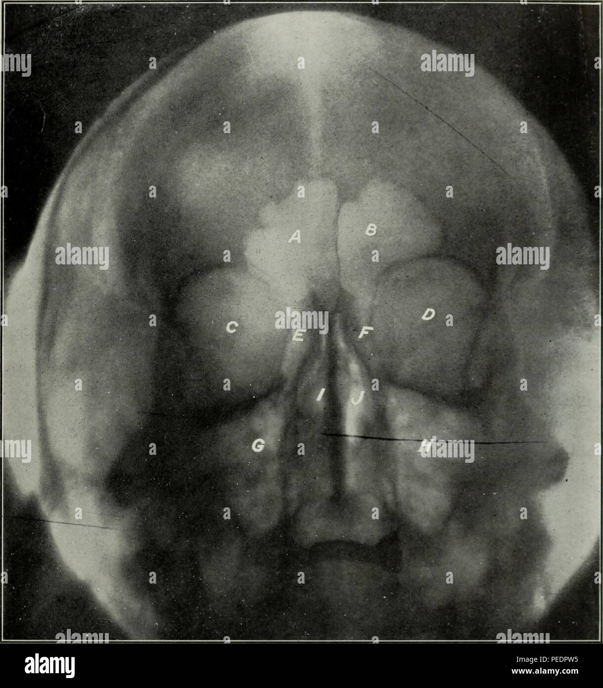


Ethmoid Sinuses High Resolution Stock Photography And Images Alamy


Brain And Face Ct Interactive Anatomy Atlas


Www Mdpi Com 75 4418 11 2 250 Pdf


Startradiology


3


Orbits Anatomy Mri Orbits And Paranasal Sinuses Anatomy Free Cross Sectional Anatomy


21 Best Paranasal Sinuses Ideas Paranasal Sinuses Sinusitis Radiology


Startradiology


Normal Ct Paranasal Sinuses Radiology Case Radiopaedia Org


Imaging Of Maxillary Sinusitis Waters View Ct Scan Mri Otolaryngology Houston
:background_color(FFFFFF):format(jpeg)/images/article/en/the-paranasal-sinuses/972PC0nYOzlz7wqSgLmNA_sinus_frontalis_large_u9Vfozc0uUoMtc6KtIaUfw.png)


Paranasal Sinuses Anatomy And Clinical Aspects Kenhub
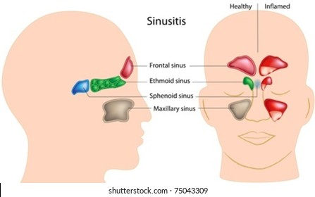


Paranasal Sinuses Images Stock Photos Vectors Shutterstock


Sphenoid Sinus Normal Anatomy Variants
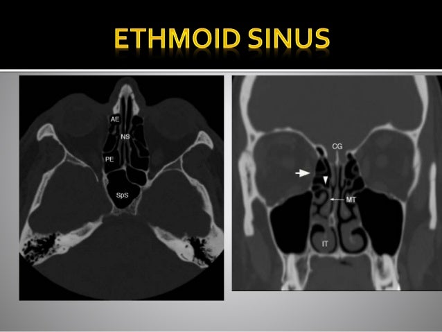


Ct Anatomy Of Para Nasal Sinuses


Www Scdentists Org Docs Librariesprovider130 Default Document Library Sinus Vs Tooth Presentation Jeffrey Adams Md578a18ddb07d6e0c8f46ff0000eea05b Pdf Sfvrsn 0


Brain And Face Ct Interactive Anatomy Atlas
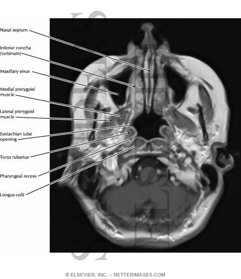


Nose And Paranasal Sinuses


D Nb Info 34


Solved Label The Grooves For Venous Sinuses In The Superior View Of The Cranial Cavity Course Hero


Improvement Diagnostic Accuracy Of Sinusitis Recognition In Paranasal Sinus X Ray Using Multiple Deep Learning Models Abstract Europe Pmc


A E Coronal Axial And Sagittal Ct Images Of The Bone Window Download Scientific Diagram
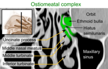


Nasal Cavity Wikipedia


コメント
コメントを投稿